Your MRI Is One of the Worst Predictors of Low Back Pain
If you’ve been told your MRI explains why your back hurts — you’re not alone.
Disc bulges. Degeneration. “Wear and tear.”
These findings sound serious, yet many people with the same scans have no pain at all.
At the same time, others experience persistent or debilitating back pain despite being told their imaging is “normal”.
This disconnect is frustrating — and confusing.
At Neurohealth Wellness on Sydney’s Northern Beaches, we see this every day. People come in with scans, reports, and unanswered questions, wondering why treatment hasn’t worked when the “problem” seems obvious on paper.
Modern pain science tells a very different story: what shows up on an MRI often has little to do with how much pain someone experiences — or how well they recover.
When Scans Don’t Tell the Whole Story: What “Abnormal” Really Means
Modern imaging has given us incredible detail into the human body. MRI and CT scans can show every curve, line, and shadow of our joints, discs, and soft tissues. But here’s the surprising truth: many of the things reported as “abnormal” on scans are found in people who have no pain at all.
At Neurohealth Wellness on Sydney’s Northern Beaches, we often see patients worried after a scan has shown a disc bulge, hip impingement, or shoulder tear. But the science tells us something important: these findings are often a normal part of being human and don’t always explain symptoms.
Spinal Discs: Ageing Gracefully
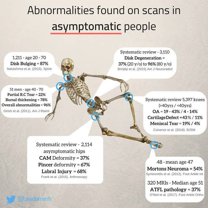
A large 2015 review showed that disc degeneration is present in 37% of 20-year-olds and climbs steadily to 96% of people in their 80s. Disc bulges were also incredibly common, appearing in up to 87% of people over 20 – many of whom had no pain at all.
More recent studies confirm the same trend. Degenerative features such as bulges, protrusions, and annular tears are frequently seen in people without back pain. Even Schmorl’s nodes – where disc material pushes into the vertebra – are found in around 15% of symptom-free adults.
In short: disc changes are not always a “problem” but often a natural part of ageing.
The Knee and Shoulder Story
Knee scans can look worrying, but research shows that cartilage defects, meniscal tears, and even early osteoarthritis changes often appear in people with no symptoms. A high-resolution MRI study found structural “abnormalities” were surprisingly common in healthy, pain-free adults.
The same applies to the shoulder. A systematic review published in 2024 reported that 30–75% of people without painstill had imaging findings such as partial rotator cuff tears or bursitis. These may sound alarming, but they don’t necessarily equal dysfunction.
Hips: Cam and Pincer Morphology
Another area where imaging often causes confusion is the hip. Cam and pincer morphologies – bony shapes linked to femoroacetabular impingement (FAI) – are frequently picked up on scans. A large study of asymptomatic people found cam morphology in around 37% of hips and even higher rates in athletes.
Interestingly, the presence of these changes doesn’t always lead to hip pain. While certain shapes can increase the risk of future problems, many people live active, pain-free lives despite having these features on MRI.
Incidental Findings: The Unexpected “Extras”
Imaging sometimes picks up unrelated findings known as “incidentalomas”. A recent review found that about 4% of people had potentially serious incidental findings, and 12% had abnormalities of uncertain significance. Importantly, only around 20% of the “serious” ones turned out to be clinically significant after follow-up.
This shows that while advanced imaging is powerful, it can also create unnecessary worry when harmless variations are labelled as disease.
We see this pattern regularly in people from Beacon Hill, Brookvale, and Dee Why who’ve been told their scan explains their pain — yet their symptoms persist because the issue lies in how the whole body and nervous system are adapting, not what the image shows.
What Actually Drives Ongoing Back Pain
Research consistently shows that persistent low back pain is influenced more by:
- Movement efficiency
- Nervous system sensitivity
- Load tolerance and recovery capacity
- Sleep, stress, and physical activity levels
Rather than being a simple “structural problem”, pain often reflects how the body is coping with cumulative stress over time.
This is where chiropractic care becomes relevant — not as a way to “fix” what’s seen on a scan, but as a way to improve how the spine, nervous system, and surrounding joints move, adapt, and tolerate load.
At Neurohealth Wellness, chiropractic assessments focus on understanding how the whole system is functioning — not just what an image shows.
Chiropractic care at Neurohealth Wellness
What This Means for You
At Neurohealth Wellness, we remind our patients of three key truths:
- Imaging is just one piece of the puzzle. A scan shows structure, not function. Pain and movement tell us far more about how your body is working.
- Abnormal doesn’t always mean unhealthy. Disc bulges, hip morphology, or rotator cuff changes are often part of normal ageing and adaptation.
- Context is everything. Clinical assessment, your history, and your goals matter more than a single line on a scan report.
The Neurohealth Approach
We focus on restoring function, movement, and balance – not just chasing scan findings. Whether it’s through chiropractic care, soft tissue therapy, acupuncture, hypnotherapy, or rehabilitation, our goal is to help you live without pain and prevent future issues, rather than worrying over every imperfection seen on a scan.
Frequently Asked Questions About MRI Scans and Back Pain
Can I still have back pain if my MRI is “normal”?
Yes. Many people experience significant pain without visible structural findings. Pain is strongly influenced by nervous system sensitivity, movement patterns, and load tolerance.
Do disc bulges always cause pain?
No. Disc bulges and degeneration are extremely common in people with no pain at all. Their presence does not automatically mean they are the source of symptoms.
If my MRI shows degeneration, does that mean my back is damaged?
Not necessarily. Degenerative changes are often part of normal ageing and do not predict pain, disability, or future outcomes on their own.
Can chiropractic help if my scan looks bad?
Chiropractic care does not treat scan findings — it focuses on improving movement, reducing nervous system overload, and helping the body tolerate load more effectively.
Why hasn’t treatment worked if the MRI shows a problem?
Treatments that focus only on structural findings may miss the broader contributors to pain, such as movement inefficiency, stress load, and recovery capacity.
A Better Way to Understand Back Pain
MRI scans can be useful in specific situations — but they are a poor predictor of pain, recovery, and long-term outcomes for most people.
Understanding back pain requires looking beyond images and addressing how the body moves, adapts, and responds to stress.
At Neurohealth Wellness, this perspective guides how we assess and support people with persistent or recurring back pain — especially those who feel stuck after being told their scan explains everything.
If you’d like to explore how this applies to your own situation, you’re welcome to book an initial consultation or learn more about our chiropractic approach.
👉 Booking link:
https://www.neurohealthwellness.com.au/booking
References
- Brinjikji, W., Luetmer, P. H., Comstock, B., Bresnahan, B. W., Chen, L. E., Deyo, R. A., ... & Jarvik, J. G. (2015). Systematic literature review of imaging features of spinal degeneration in asymptomatic populations. American Journal of Neuroradiology, 36(4), 811–816.
- Nakashima, H., Yukawa, Y., Suda, K., Yamagata, M., Ueta, T., & Kato, F. (2015). Abnormal findings on magnetic resonance images of the cervical spines in 1211 asymptomatic subjects. Spine, 40(6), 392–398.
- Culvenor, A. G., Øiestad, B. E., Hart, H. F., Stefanik, J. J., Guermazi, A., Crossley, K. M. (2019). Prevalence of knee osteoarthritis features on MRI in asymptomatic uninjured adults: a systematic review and meta-analysis. British Journal of Sports Medicine, 53(20), 1268–1278.
- Schleich, C., et al. (2020). MRI abnormalities in asymptomatic knees using a 3T system with a dedicated multichannel coil: prevalence and distribution of findings. Skeletal Radiology, 49(8), 1281–1290.
- Simel, D. L., et al. (2024). Prevalence of shoulder abnormalities in asymptomatic adults: a systematic review. Journal of Shoulder and Elbow Surgery, 33(6), 1234–1245.
- Frank, J. M., Harris, J. D., Erickson, B. J., Slikker, W., Bush-Joseph, C. A., Salata, M. J., & Nho, S. J. (2015). Prevalence of femoroacetabular impingement imaging findings in asymptomatic volunteers: a systematic review. Arthroscopy, 31(6), 1199–1204.
- Agricola, R., et al. (2013). Cam impingement of the hip: a risk factor for hip osteoarthritis. Nature Reviews Rheumatology, 9(10), 630–634.
- Morris, Z., Whiteley, W. N., Longstreth, W. T., Weber, F., Lee, Y. C., Tsushima, Y., ... & Wardlaw, J. M. (2009). Incidental findings on brain magnetic resonance imaging: systematic review and meta-analysis. BMJ, 339, b3016.
- Lumbreras, B., Donat, L., & Hernández-Aguado, I. (2010). Incidental findings in imaging diagnostic tests: a systematic review. British Journal of Radiology, 83(988), 276–289.

